Copyright
©2007 Baishideng Publishing Group Co.
World J Gastroenterol. Jan 28, 2007; 13(4): 639-642
Published online Jan 28, 2007. doi: 10.3748/wjg.v13.i4.639
Published online Jan 28, 2007. doi: 10.3748/wjg.v13.i4.639
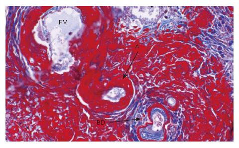
Figure 3 A smaller portal tract.
Fibrinoid material is deposited in the connective tissue, walls of the arteries and periductular connective tissue (Masson's trichrome x 20). PV: portal vein, A: artery, BD: bile duct.
- Citation: Hano H, Takagi I, Nagatsuma K, Lu T, Meng C, Chiba S. An autopsy case showing massive fibrinoid necrosis of the portal tracts of the liver with cholangiographic findings similar to those of primary sclerosing cholangitis. World J Gastroenterol 2007; 13(4): 639-642
- URL: https://www.wjgnet.com/1007-9327/full/v13/i4/639.htm
- DOI: https://dx.doi.org/10.3748/wjg.v13.i4.639


