Copyright
©2007 Baishideng Publishing Group Inc.
World J Gastroenterol. Oct 21, 2007; 13(39): 5217-5225
Published online Oct 21, 2007. doi: 10.3748/wjg.v13.i39.5217
Published online Oct 21, 2007. doi: 10.3748/wjg.v13.i39.5217
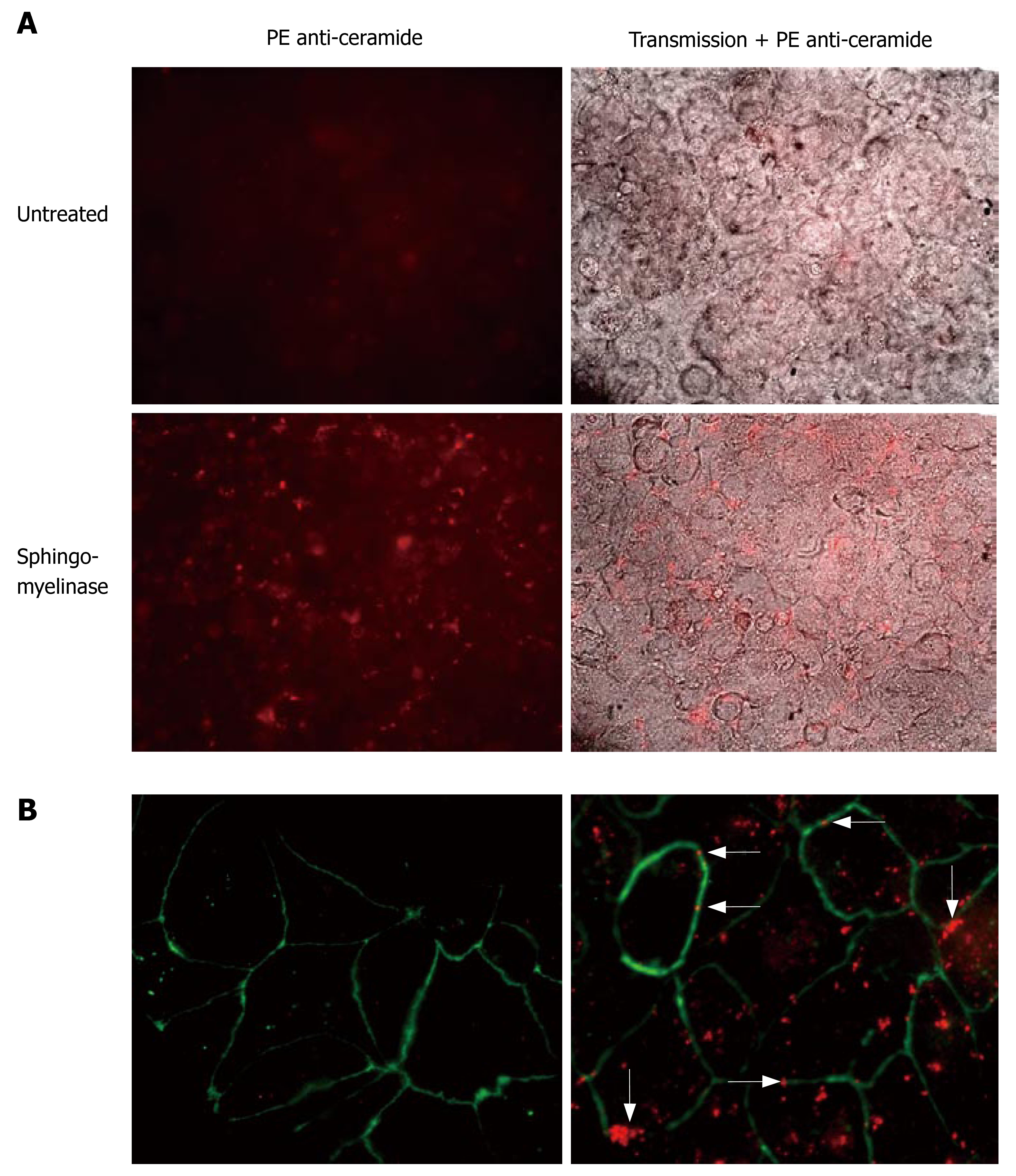
Figure 5 Ceramide clusters at the sites of cell-cell contacts.
Caco-2 cell layers were incubated with 0.25 U/mL SMase for 10 min or left untreated. After fixation of the cells with paraformaldehyde, ceramide was visualized by staining of the cells with the monoclonal anti-ceramide 15B4 antibody and PE-coupled anti-mouse antibodies. A: Top, left: Non-stimulated cells displayed a certain level of background staining that was used to determine the time of exposure. Top, right: Overlay of fluorescence and transmission. Bottom, left: Incubation with SMase resulted in formation of multiple ceramide-clusters. Bottom, right: Overlay of fluorescence and transmission. Close inspection of the clusters revealed a frequent localization of the clusters at the sites of cell/cell-contact; B: Costaining of tight junctions with FITC-labeled anti ZO-1 Abs. Left: non-stimulated. Right: Predominant colocalization of ceramide-clusters with tight junctions upon stimulation with SMase (arrows indicate ceramide-clusters at the sites of cell-cell contact).
- Citation: Bock J, Liebisch G, Schweimer J, Schmitz G, Rogler G. Exogenous sphingomyelinase causes impaired intestinal epithelial barrier function. World J Gastroenterol 2007; 13(39): 5217-5225
- URL: https://www.wjgnet.com/1007-9327/full/v13/i39/5217.htm
- DOI: https://dx.doi.org/10.3748/wjg.v13.i39.5217


