Copyright
©2007 Baishideng Publishing Group Inc.
World J Gastroenterol. Oct 21, 2007; 13(39): 5208-5216
Published online Oct 21, 2007. doi: 10.3748/wjg.v13.i39.5208
Published online Oct 21, 2007. doi: 10.3748/wjg.v13.i39.5208
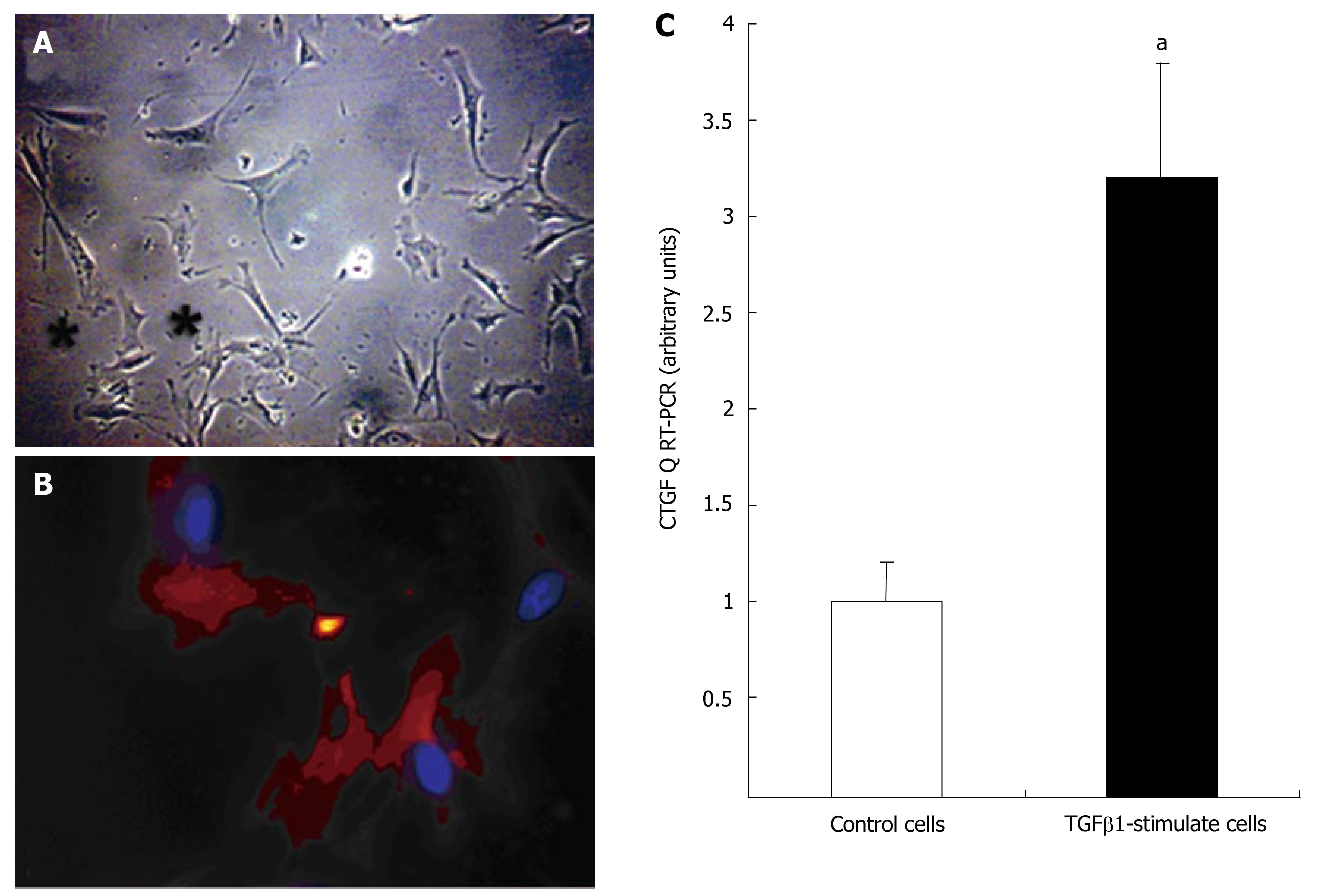
Figure 4 Micrographs of primary cultured human myofibroblasts isolated from human fibrotic material (SI carcinoid tumor).
A: Light microscopy identified the typical stellate shape (black stars) in 5-day cultured cells (200 × magnification); B: Immunostaining with α-smooth muscle actin (Cy-5-red stain; nuclei are blue-DAPI) in same cells after 7-d culture (x 600); C: Message levels of CTGF determined by Q RT-PCR in primary cultured human myofibroblasts. CTGF was significantly over-expressed (about 3-fold) in TGFβ1 (10-7 mol/L, 24 h) stimulated cells compared to control (un-stimulated) cells (aP < 0.05), mean ± SE, n = 3.
- Citation: Kidd M, Modlin I, Shapiro M, Camp R, Mane S, Usinger W, Murren J. CTGF, intestinal stellate cells and carcinoid fibrogenesis. World J Gastroenterol 2007; 13(39): 5208-5216
- URL: https://www.wjgnet.com/1007-9327/full/v13/i39/5208.htm
- DOI: https://dx.doi.org/10.3748/wjg.v13.i39.5208


