Copyright
©2007 Baishideng Publishing Group Inc.
World J Gastroenterol. Oct 21, 2007; 13(39): 5208-5216
Published online Oct 21, 2007. doi: 10.3748/wjg.v13.i39.5208
Published online Oct 21, 2007. doi: 10.3748/wjg.v13.i39.5208
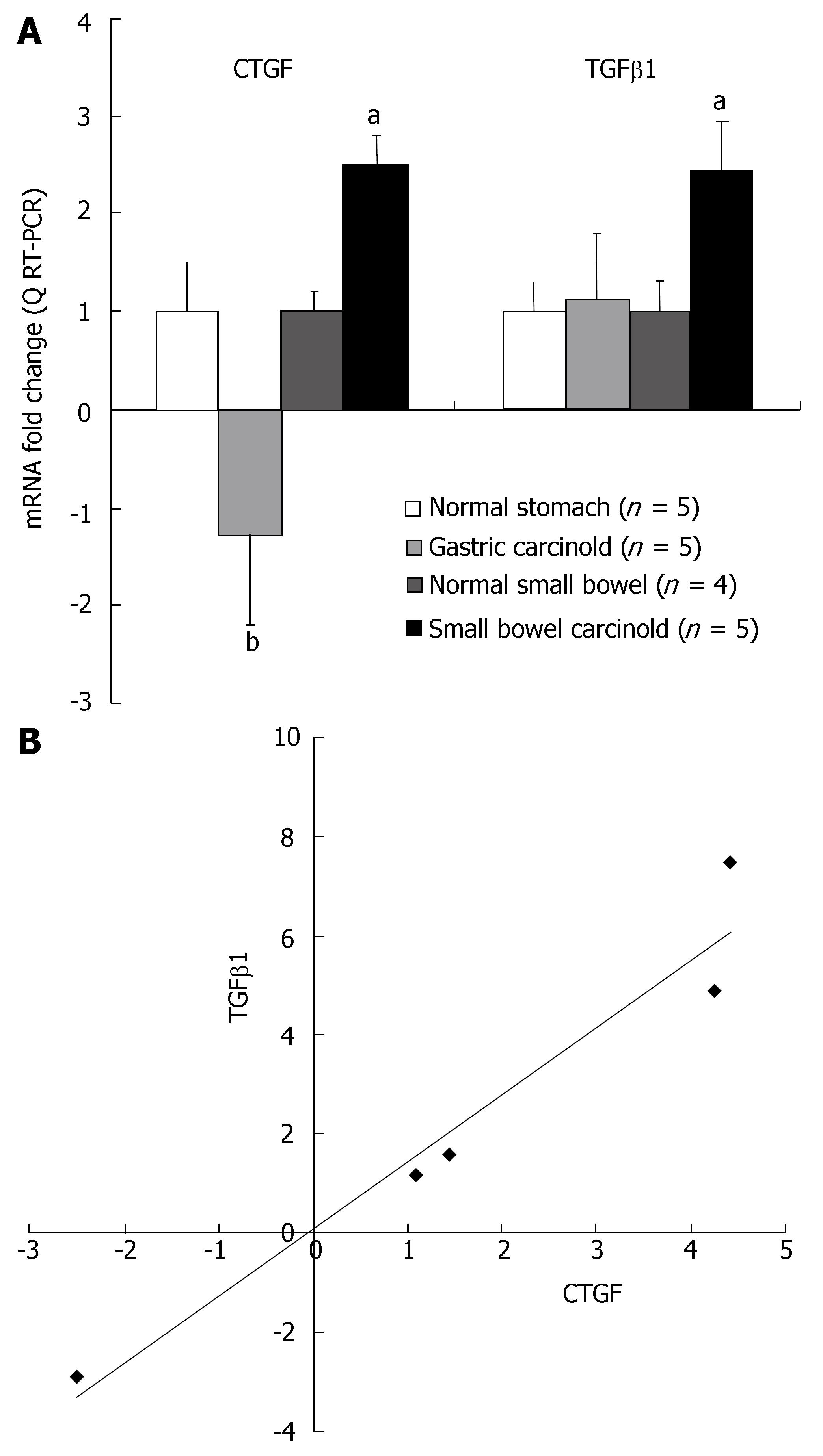
Figure 1 A: Message levels of both CTGF and TGFβ1 determined by Q RT-PCR.
Levels were corrected against expression of the housekeeping gene, GAPDH, compared to similarly corrected gene levels in normal mucosa, and represented as fold increase over normal (1.0). TGFβ1 was significantly over-expressed (about 2.5-fold) in SI carcinoid tumor samples compared to normal mucosa (aP < 0.05) but not the gastric carcinoids. CTGF was significantly over-expressed (about 2.5-fold) in SI carcinoid tumor samples compared to normal mucosa (aP < 0.05) while gastric carcinoids had significantly decreased CTGF compared to SI carcinoid tumors (bP < 0.01). Mean ± SE; B: Correlation analysis of QRT-PCR results in SI EC cell carcinoid tumors. There was a good correlation between CTGF and TGFβ1 transcript levels in tumor samples (R2 = 0.9445, P < 0.01, n = 5).
- Citation: Kidd M, Modlin I, Shapiro M, Camp R, Mane S, Usinger W, Murren J. CTGF, intestinal stellate cells and carcinoid fibrogenesis. World J Gastroenterol 2007; 13(39): 5208-5216
- URL: https://www.wjgnet.com/1007-9327/full/v13/i39/5208.htm
- DOI: https://dx.doi.org/10.3748/wjg.v13.i39.5208


