Copyright
©2007 Baishideng Publishing Group Co.
World J Gastroenterol. Oct 14, 2007; 13(38): 5139-5146
Published online Oct 14, 2007. doi: 10.3748/wjg.v13.i38.5139
Published online Oct 14, 2007. doi: 10.3748/wjg.v13.i38.5139
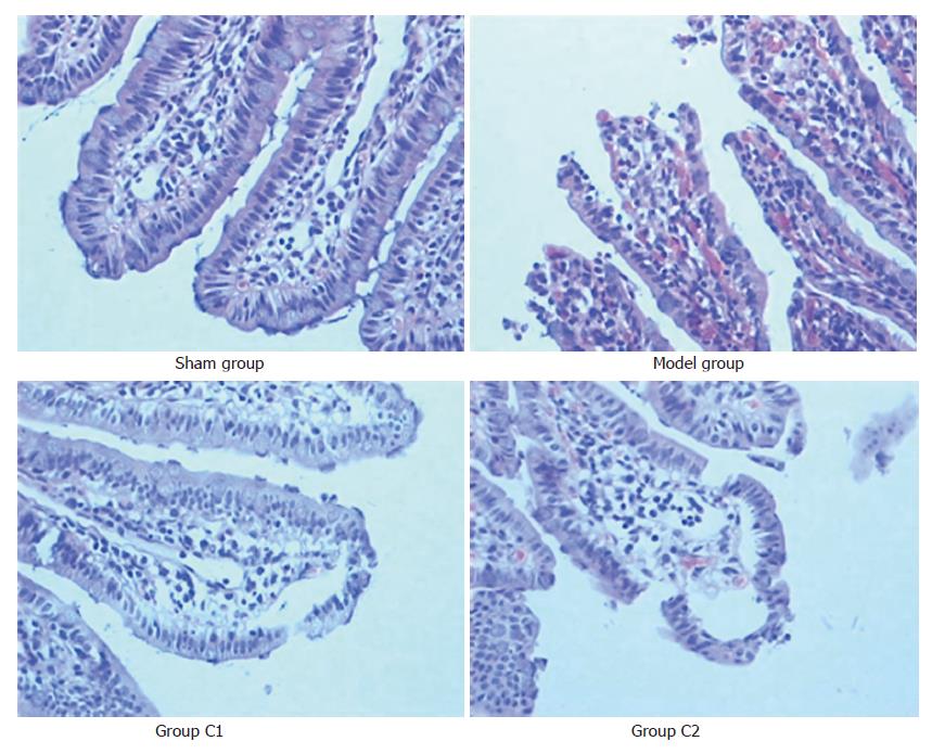
Figure 1 Microscopic appearance after hematoxylin and eosin staining (× 200).
In the model group, there are multiple erosions and bleeding in mucosal epithelial layer; the villus and glands were normal and no inflammatory cell infiltration was observed in mucosal epithelial layer in sham group; these mucosal changes are ameliorated by treatment with Cromolyn Sodium (group C1 and group C2).
- Citation: Hei ZQ, Gan XL, Luo GJ, Li SR, Cai J. Pretreatment of cromolyn sodium prior to reperfusion attenuates early reperfusion injury after the small intestine ischemia in rats. World J Gastroenterol 2007; 13(38): 5139-5146
- URL: https://www.wjgnet.com/1007-9327/full/v13/i38/5139.htm
- DOI: https://dx.doi.org/10.3748/wjg.v13.i38.5139


