Copyright
©2007 Baishideng Publishing Group Co.
World J Gastroenterol. Oct 7, 2007; 13(37): 4986-4995
Published online Oct 7, 2007. doi: 10.3748/wjg.v13.i37.4986
Published online Oct 7, 2007. doi: 10.3748/wjg.v13.i37.4986
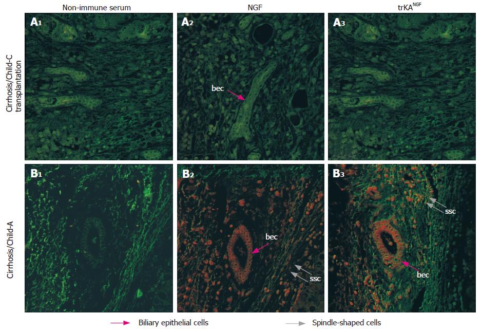
Figure 3 NGF and trkANGF distribution in cirrhotic tissues by confocal microscopy.
A1: Liver specimens obtained before transplantation from patients with cirrhosis but without HCC (Child-Pugh C); A2: NGF distribution; A3: trkANGF distribution; B1: Specimens from patients with cirrhosis, also suffering from HCC, at early staging (Child-Pugh A); B2: NGF distribution; B3: trkANGF distribution. NGF or trkANGF immunoreaction (red hue) is particularly evident on biliary epithelial cells and on spindle-shaped cells, in cirrhotic tissue from patient at early staging (Child-Pugh A), but in presence of HCC, while no immunoreaction is observed in liver specimens resected before transplantation (Child-Pugh C) from patients without HCC. No immunoreaction is observed in controls performed by substitution of primary antisera with non-immune rabbit serum. bec: biliary epithelial cells; ssc: spindle-shaped cells.
- Citation: Rasi G, Serafino A, Bellis L, Lonardo MT, Andreola F, Zonfrillo M, Vennarecci G, Pierimarchi P, Vallebona PS, Ettorre GM, Santoro E, Puoti C. Nerve growth factor involvement in liver cirrhosis and hepatocellular carcinoma. World J Gastroenterol 2007; 13(37): 4986-4995
- URL: https://www.wjgnet.com/1007-9327/full/v13/i37/4986.htm
- DOI: https://dx.doi.org/10.3748/wjg.v13.i37.4986


