Copyright
©2007 Baishideng Publishing Group Co.
World J Gastroenterol. Sep 28, 2007; 13(36): 4897-4902
Published online Sep 28, 2007. doi: 10.3748/wjg.v13.i36.4897
Published online Sep 28, 2007. doi: 10.3748/wjg.v13.i36.4897
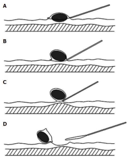
Figure 1 Schematic diagram for “pushing” technique.
A: The snare was placed around the leiomyoma; B: The head (gastroscopy) or anal (colonoscopy) side of leiomyoma was pushed by the insulated cannula of snare to form a semipedunculation; C: The snare was tightened gradually and total leiomyoma was captured; D: The leiomyoma was resected completely.
- Citation: Zhou XD, Lv NH, Chen HX, Wang CW, Zhu X, Xu P, Chen YX. Endoscopic management of gastrointestinal smooth muscle tumor. World J Gastroenterol 2007; 13(36): 4897-4902
- URL: https://www.wjgnet.com/1007-9327/full/v13/i36/4897.htm
- DOI: https://dx.doi.org/10.3748/wjg.v13.i36.4897


