Copyright
©2007 Baishideng Publishing Group Co.
World J Gastroenterol. Sep 28, 2007; 13(36): 4891-4896
Published online Sep 28, 2007. doi: 10.3748/wjg.v13.i36.4891
Published online Sep 28, 2007. doi: 10.3748/wjg.v13.i36.4891
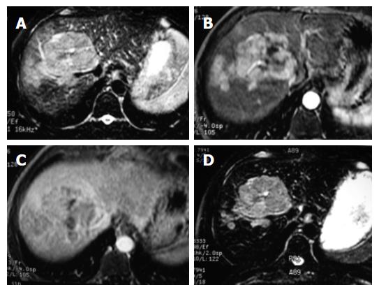
Figure 3 HCC image.
A: The lesion appears hyperintense on the pre-contrast T2WI FSE image, but is not clearly delineated; B: The lesion is markedly enhanced during the hepatic arterial phase; C: The lesion appears hypointence during the portal venous phase, but is not clearly delineated; D: The main lesion and satellite nodules are clearly delineated from the surrounding hypointense liver parenchyma on the ferucarbotran-enhanced T2WI image during the accumulation phase.
-
Citation: Cheng WZ, Zeng MS, Yan FH, Rao SX, Shen JZ, Chen CZ, Zhang SJ, Shi WB. Ferucarbotran
versus Gd-DTPA-enhanced MR imaging in the detection of focal hepatic lesions. World J Gastroenterol 2007; 13(36): 4891-4896 - URL: https://www.wjgnet.com/1007-9327/full/v13/i36/4891.htm
- DOI: https://dx.doi.org/10.3748/wjg.v13.i36.4891


