Copyright
©2007 Baishideng Publishing Group Co.
World J Gastroenterol. Sep 7, 2007; 13(33): 4526-4528
Published online Sep 7, 2007. doi: 10.3748/wjg.v13.i33.4526
Published online Sep 7, 2007. doi: 10.3748/wjg.v13.i33.4526
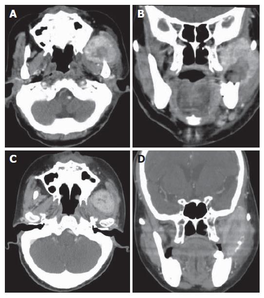
Figure 1 Axial and coronal CT scan of the neck with intravenous contrast showing a 6.
2 cm х 5.0 cm, heterogeneously enhancing mass, which appears to be a left parapharyngeal mass involving the pterygoid muscle and temporal muscle (A and B); axial and coronal CT scan of the head and neck showing a 4.0 cm х 2.5 cm mass which shrunk after completion of radiotherapy (C and D).
- Citation: Huang SF, Wu RC, Chang JTC, Chan SC, Liao CT, Chen IH, Yeh CN. Intractable bleeding from solitary mandibular metastasis of hepatocellular carcinoma. World J Gastroenterol 2007; 13(33): 4526-4528
- URL: https://www.wjgnet.com/1007-9327/full/v13/i33/4526.htm
- DOI: https://dx.doi.org/10.3748/wjg.v13.i33.4526


