Copyright
©2007 Baishideng Publishing Group Co.
World J Gastroenterol. Sep 7, 2007; 13(33): 4520-4522
Published online Sep 7, 2007. doi: 10.3748/wjg.v13.i33.4520
Published online Sep 7, 2007. doi: 10.3748/wjg.v13.i33.4520
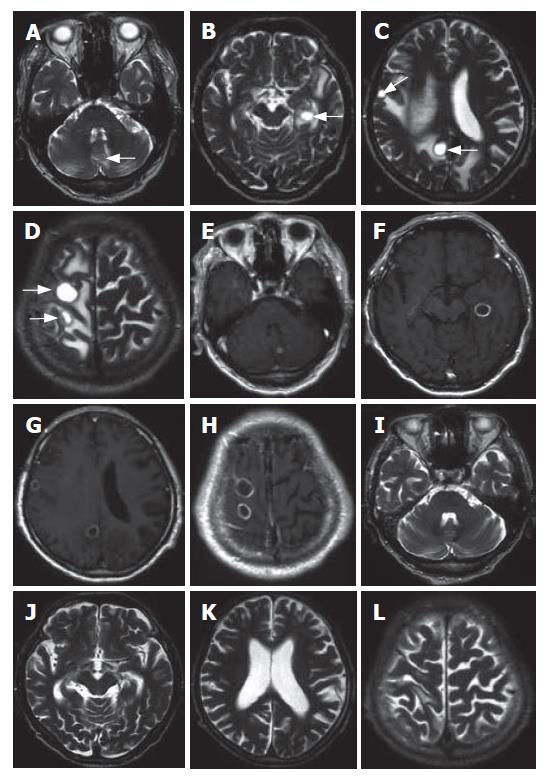
Figure 3 Unenhanced T2-weighted MR images show hyperintense tumors in the cerebrum and cerebellum (arrows) (A, B, C, D).
Contrast-enhanced T1-weighted MR images show ring-shaped enhancement of the tumors (E, F, G, H). On T2-weighted, unenhanced MR images, captured 3 mo after the completion of radiotherapy, the brain tumors have almost completely disappeared (I, J, K, L).
- Citation: Toshikuni N, Morii K, Yamamoto M. Radiotherapy for multiple brain metastases from hepatocellular carcinomas. World J Gastroenterol 2007; 13(33): 4520-4522
- URL: https://www.wjgnet.com/1007-9327/full/v13/i33/4520.htm
- DOI: https://dx.doi.org/10.3748/wjg.v13.i33.4520


