Copyright
©2007 Baishideng Publishing Group Co.
World J Gastroenterol. Sep 7, 2007; 13(33): 4498-4503
Published online Sep 7, 2007. doi: 10.3748/wjg.v13.i33.4498
Published online Sep 7, 2007. doi: 10.3748/wjg.v13.i33.4498
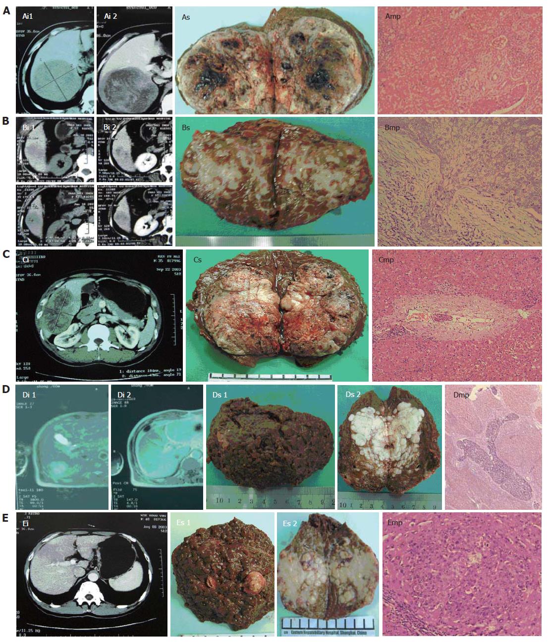
Figure 1 Imaging (i), specimens (s) and microscopic pathology (mp) of 5 PLC patients.
A: case 24, tumor size 12.0 cm x 11.0 cm x 10.5cm, with microscopic tumor thrombi (metastatic distance 6 mm, x 100); B: case 40, tumor size 4.0 cm x 3.8 cm x 3.5 cm, with microscopic capsular infiltration (x 200); C: case 58, tumor size 10.5 cm x 6.5 cm x 6.0 cm, with microscopic tumor thrombi (metastatic distance 2 mm, x 100); D: case 38, tumor size 6.5 cm x 4.0 cm x 3.6 cm, with macroscopic tumor thrombi in the branches of right portal vein and microscopic tumor thrombi of arborization (x 16); E: case 52, tumor size 7.0 cm x 5.0 cm x 5.0 cm, with macroscopic tumor thrombi in the branches of right portal vein and microsatellites (metastatic distance 3.5 mm, x 100).
- Citation: Zhou XP, Quan ZW, Cong WM, Yang N, Zhang HB, Zhang SH, Yang GS. Micrometastasis in surrounding liver and the minimal length of resection margin of primary liver cancer. World J Gastroenterol 2007; 13(33): 4498-4503
- URL: https://www.wjgnet.com/1007-9327/full/v13/i33/4498.htm
- DOI: https://dx.doi.org/10.3748/wjg.v13.i33.4498


