Copyright
©2007 Baishideng Publishing Group Co.
World J Gastroenterol. Aug 21, 2007; 13(31): 4177-4184
Published online Aug 21, 2007. doi: 10.3748/wjg.v13.i31.4177
Published online Aug 21, 2007. doi: 10.3748/wjg.v13.i31.4177
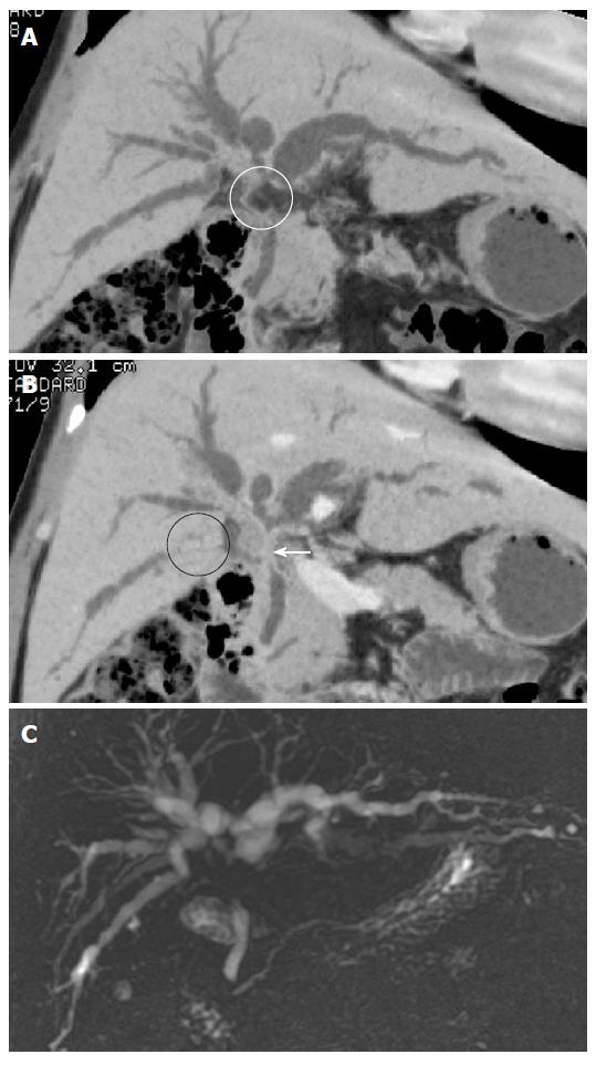
Figure 12 Klatskin’s tumor in a 65 years old man with jaundice.
A: MinIP (coronal oblique 18.8 mm thickness slab) image shows focal disruption (white circle) of the common hepatic duct due to the adjacent perihepatic fat; B: MinIP (coronal oblique 6.2 mm thickness slab) image shows focal loss (black circle) of the right posterior inferior segmental duct due to exclusion by out of the slab range. However, severe narrowing of the common hepatic duct (white arrow) is better visualized than that seen on (A); C: MRCP shows a typical Klatskin tumor with narrowing of the confluent portion of both intrahepatic bile ducts.
- Citation: Kim HC, Yang DM, Jin W, Ryu CW, Ryu JK, Park SI, Park SJ, Shin HC, Kim IY. Multiplanar reformations and minimum intensity projections using multi-detector row CT for assessing anomalies and disorders of the pancreaticobiliary tree. World J Gastroenterol 2007; 13(31): 4177-4184
- URL: https://www.wjgnet.com/1007-9327/full/v13/i31/4177.htm
- DOI: https://dx.doi.org/10.3748/wjg.v13.i31.4177


