Copyright
©2007 Baishideng Publishing Group Co.
World J Gastroenterol. Aug 7, 2007; 13(29): 3939-3947
Published online Aug 7, 2007. doi: 10.3748/wjg.v13.i29.3939
Published online Aug 7, 2007. doi: 10.3748/wjg.v13.i29.3939
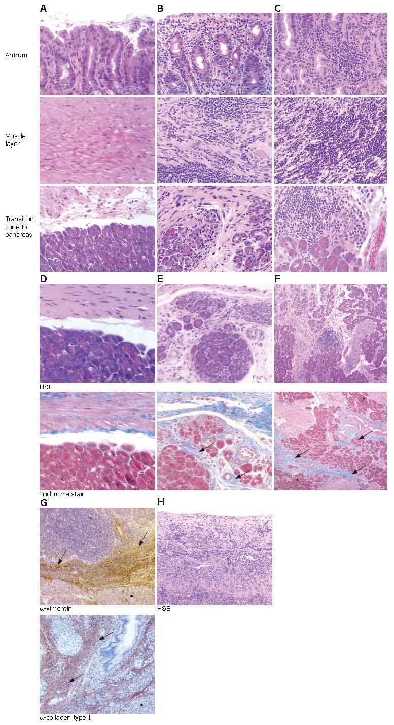
Figure 1 Histopathological changes in the gastric mucosa and adjacent pancreatic tissue of Mongolian gerbils seven months after inoculation.
(HE stain, × 40). A: Control group, no inflammatory cell infiltrates and pathological changes; B: H pylori B128 WT-strain infected gerbils, showed severe inflammatory cell infiltrates in the antral mucosa extending into the muscularis externa, and chronic pancreatitis; C: H pylori B128 ΔcagY-mutant infected gerbils, revealed a severe antral gastritis extending into the muscularis externa, and chronic pancreatitis; D: Control groups, no pancreatic injury; E: Chronic pancreatitis with marked atrophy and fibrous replacement of pancreatic acini was highlighted by trichrome staining of WT-infected gerbils and F: In gerbils infected with ΔcagY-mutant. G: Fibrous tissue in the pancreas of infected Mongolian gerbils detected with anti-vimentin and anti-collagen typeIantibody; H: Severe ulceration of antral mucosa with marked inflammatory cell infiltration into submucosa and transmural inflammation into adjacent pancreatic lobules in WT-infected gerbil. Asterisks indicate pancreatic acini while arrows mark the fibrotic changes within the pancreas.
-
Citation: Rieder G, Karnholz A, Stoeckelhuber M, Merchant JL, Haas R.
H pylori infection causes chronic pancreatitis in Mongolian gerbils. World J Gastroenterol 2007; 13(29): 3939-3947 - URL: https://www.wjgnet.com/1007-9327/full/v13/i29/3939.htm
- DOI: https://dx.doi.org/10.3748/wjg.v13.i29.3939


