Copyright
©2007 Baishideng Publishing Group Co.
World J Gastroenterol. Jul 28, 2007; 13(28): 3892-3894
Published online Jul 28, 2007. doi: 10.3748/wjg.v13.i28.3892
Published online Jul 28, 2007. doi: 10.3748/wjg.v13.i28.3892
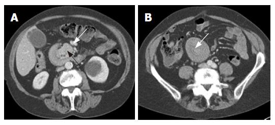
Figure 1 Enhanced CT showing the ampullary mass (black arrow) invaginating the duodenum (A) and the duodenoduodenal intussusception in the midline in the infracolic compartment (B).
- Citation: Gardner-Thorpe J, Hardwick R, Carroll N, Gibbs P, Jamieson N, Praseedom R. Adult duodenal intussusception associated with congenital malrotation. World J Gastroenterol 2007; 13(28): 3892-3894
- URL: https://www.wjgnet.com/1007-9327/full/v13/i28/3892.htm
- DOI: https://dx.doi.org/10.3748/wjg.v13.i28.3892


