Copyright
©2007 Baishideng Publishing Group Co.
World J Gastroenterol. Jul 21, 2007; 13(27): 3760-3762
Published online Jul 21, 2007. doi: 10.3748/wjg.v13.i27.3760
Published online Jul 21, 2007. doi: 10.3748/wjg.v13.i27.3760
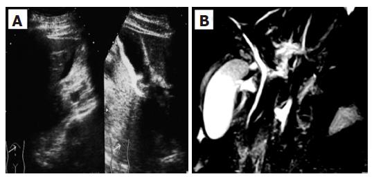
Figure 1 A: Abdominal ultrasonography (US) revealed a marked gall bladder swelling and wall thickness without stones or debris, indicating an acute cholecystitis; B: Magnetic resonance cholangiopancreatography (MRCP) revealed a similar gall bladder thickness as well as free space around the gall bladder, suggesting exudate associated with severe inflammation.
-
Citation: Kaji K, Yoshiji H, Yoshikawa M, Yamazaki M, Ikenaka Y, Noguchi R, Sawai M, Ishikawa M, Mashitani T, Kitade M, Kawaratani H, Uemura M, Yamao J, Fujimoto M, Mitoro A, Toyohara M, Yoshida M, Fukui H. Eosinophilic cholecystitis along with pericarditis caused by
Ascaris lumbricoides : A case report. World J Gastroenterol 2007; 13(27): 3760-3762 - URL: https://www.wjgnet.com/1007-9327/full/v13/i27/3760.htm
- DOI: https://dx.doi.org/10.3748/wjg.v13.i27.3760


