Copyright
©2007 Baishideng Publishing Group Inc.
World J Gastroenterol. Jul 14, 2007; 13(26): 3598-3604
Published online Jul 14, 2007. doi: 10.3748/wjg.v13.i26.3598
Published online Jul 14, 2007. doi: 10.3748/wjg.v13.i26.3598
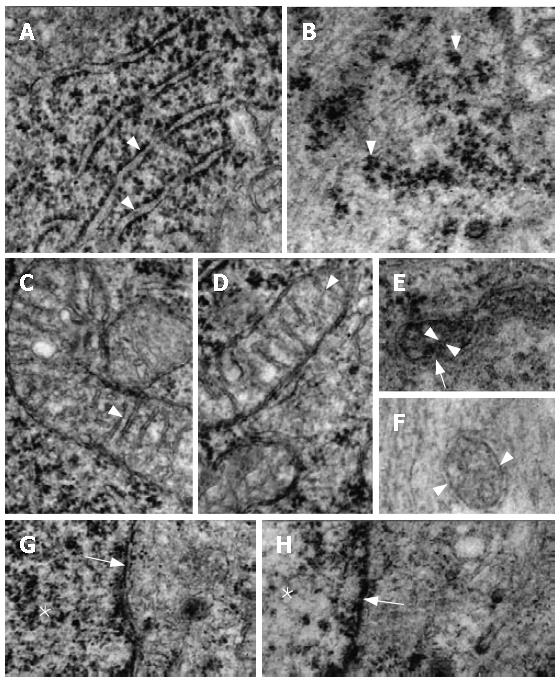
Figure 4 Electron micrographs of myenteric neurons.
A: Granular reticulum of N with the ribosomes aligned on the outer surface of the regularly-arranged membrane (arrowheads); B: Characteristic disposition of the ribosomes in clusters in D animals (arrowheads); C, D: Normal aspects of the mitochondria of N with typical transversal cristae (arrowheads); E: A electron-dense mitochondrium of N with normal double membrane (arrow) and a obliquely-oriented crista (arrowheads); F: Transversal section of a mitochondrium of D. Note the double membrane preserved (arrowheads); G: Nuclear chromatin (*) and nuclear membrane (arrow) of N; H: The same aspects in a D neuron. Compare the distribution of the nuclear chromatin (*) with a N specimen (G). (A-D: x 40000; E, F: x 60000; G and H: x 30000).
- Citation: Liberti EA, Fontes RB, Fuggi VM, Maifrino LB, Souza RR. Effects of combined pre- and post-natal protein deprivation on the myenteric plexus of the esophagus of weanling rats: A histochemical, quantitative and ultrastructural study. World J Gastroenterol 2007; 13(26): 3598-3604
- URL: https://www.wjgnet.com/1007-9327/full/v13/i26/3598.htm
- DOI: https://dx.doi.org/10.3748/wjg.v13.i26.3598


