Copyright
©2007 Baishideng Publishing Group Inc.
World J Gastroenterol. Jul 14, 2007; 13(26): 3598-3604
Published online Jul 14, 2007. doi: 10.3748/wjg.v13.i26.3598
Published online Jul 14, 2007. doi: 10.3748/wjg.v13.i26.3598
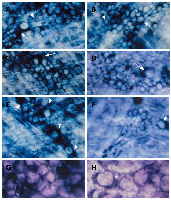
Figure 1 Myenteric neurons stained for NADH diaphorase in esophagi taken from N (A, C, E, G) and D (B, D, F, H) rats.
In both groups small (arrowheads) and large (arrows) neurons are evident (A, B). Sometimes large neurons were only weakly stained, such as in C and D (arrows) and G and H (*). the weakness of staining in large neurons; E, F: On the other hand, in N animals (E) large and intensely reactive neurons predominated (arrowheads), but their number was largely reduced inside D ganglia (F, arrowhead) (A-F: x 340; G and H: x 715).
- Citation: Liberti EA, Fontes RB, Fuggi VM, Maifrino LB, Souza RR. Effects of combined pre- and post-natal protein deprivation on the myenteric plexus of the esophagus of weanling rats: A histochemical, quantitative and ultrastructural study. World J Gastroenterol 2007; 13(26): 3598-3604
- URL: https://www.wjgnet.com/1007-9327/full/v13/i26/3598.htm
- DOI: https://dx.doi.org/10.3748/wjg.v13.i26.3598


