Copyright
©2007 Baishideng Publishing Group Inc.
World J Gastroenterol. Jul 14, 2007; 13(26): 3592-3597
Published online Jul 14, 2007. doi: 10.3748/wjg.v13.i26.3592
Published online Jul 14, 2007. doi: 10.3748/wjg.v13.i26.3592
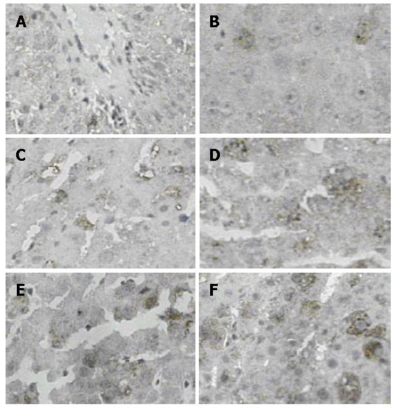
Figure 6 Immunohistochemical staining of LYZ-positive cells in rat liver.
The different staining intensity LYZ positive cells were observed in the cytoplasm of NC, SO and PH groups, surrounding the central vein(CV) and portal triad(PT). A: LYZ positive cells in NC group; B: LYZ positive cells in SO group at 6 h; C: LYZ positive cells in PH group at 6 h ; D: LYZ positive cells in PH group at 12 h; E: LYZ positive cells in PH group at 24 h ; F: LYZ positive cells in PH group at 48 h (SABC, × 200).
- Citation: Xu CP, Liu J, Liu JC, Han DW, Zhang Y, Zhao YC. Dynamic changes and mechanism of intestinal endotoxemia in partially hepatectomized rats. World J Gastroenterol 2007; 13(26): 3592-3597
- URL: https://www.wjgnet.com/1007-9327/full/v13/i26/3592.htm
- DOI: https://dx.doi.org/10.3748/wjg.v13.i26.3592


