Copyright
©2007 Baishideng Publishing Group Inc.
World J Gastroenterol. Jul 7, 2007; 13(25): 3472-3477
Published online Jul 7, 2007. doi: 10.3748/wjg.v13.i25.3472
Published online Jul 7, 2007. doi: 10.3748/wjg.v13.i25.3472
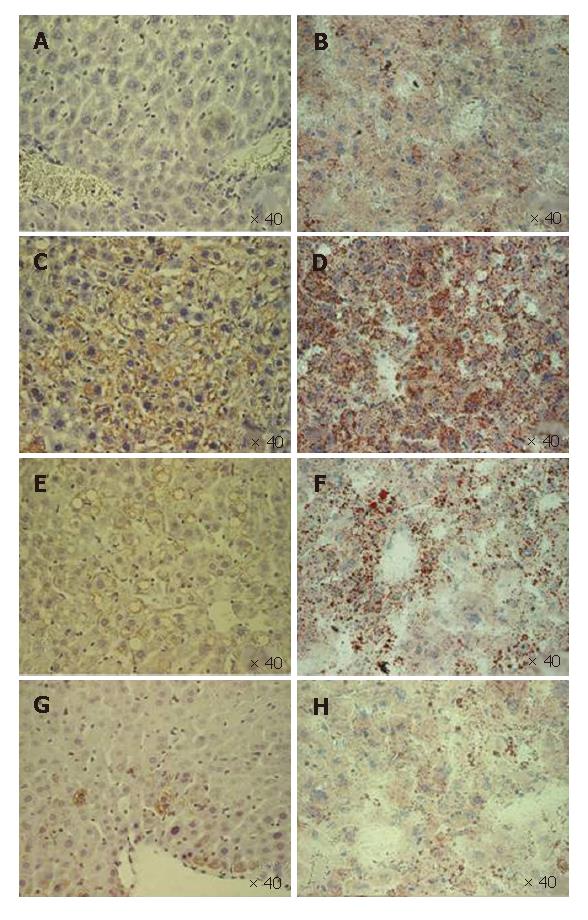
Figure 4 Immunostaining for core protein in core-expressing mice (DTM) livers fixed in 4% PFA (A, C, E and G) and the corresponding oil-red-O stain for fat in the livers of the frozen section (B, D, F and H).
The livers were from the mice on doxycycline (A and B) and those not on doxycycline (C and D) at an age of 2 mo, 4 mo (E and F) and 6 mo (G and H). The core expression parallels the degree of hepatic steatosis. Both peaked at the age of 2 mo (C and D) when the hepatic steatosis was microvesicular (D), and diminished gradually (E and H). Macrovesicular hepatic steatosis (E and F) replaced microvesicluar steatosis during evolution.
-
Citation: Chang ML, Chen JC, Yeh CT, Sheen IS, Tai DI, Chang MY, Chiu CT, Lin DY, Bissell DM. Topological and evolutional relationships between HCV core protein and hepatic lipid vesicles: Studies
in vitro and in conditionally transgenic mice. World J Gastroenterol 2007; 13(25): 3472-3477 - URL: https://www.wjgnet.com/1007-9327/full/v13/i25/3472.htm
- DOI: https://dx.doi.org/10.3748/wjg.v13.i25.3472


