Copyright
©2007 Baishideng Publishing Group Inc.
World J Gastroenterol. Jul 7, 2007; 13(25): 3472-3477
Published online Jul 7, 2007. doi: 10.3748/wjg.v13.i25.3472
Published online Jul 7, 2007. doi: 10.3748/wjg.v13.i25.3472
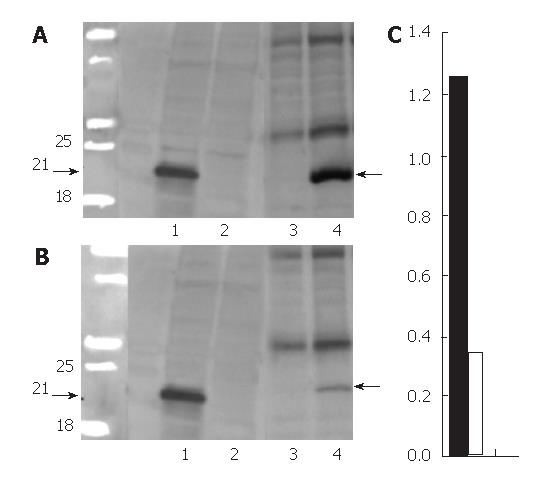
Figure 1 Western blotting for protein extracted from transfected HeLa cells and representative transgenic mice liver.
A: two months old mice; B: the faint 21-KD-core band (line 4) in six months old mice. Lanes 1 and 2: proteins from transfected HeLa cells without and with the administration of doxycycline which served as positive and negative controls. Lanes 3 and 4: proteins from the transgenic mice fed with and without doxycycline chow, respectively; C: The quantitative comparison of the blotting. Black bar, female 2 mo old DTM liver protein (1.26 ± 0.08); white bar: female 6 mo old DTM liver protein (0.34 ± 0.05), P = 0.002 between black and white bars (mean ± SD, n = 3).
-
Citation: Chang ML, Chen JC, Yeh CT, Sheen IS, Tai DI, Chang MY, Chiu CT, Lin DY, Bissell DM. Topological and evolutional relationships between HCV core protein and hepatic lipid vesicles: Studies
in vitro and in conditionally transgenic mice. World J Gastroenterol 2007; 13(25): 3472-3477 - URL: https://www.wjgnet.com/1007-9327/full/v13/i25/3472.htm
- DOI: https://dx.doi.org/10.3748/wjg.v13.i25.3472


