Copyright
©2007 Baishideng Publishing Group Inc.
World J Gastroenterol. Jul 7, 2007; 13(25): 3409-3416
Published online Jul 7, 2007. doi: 10.3748/wjg.v13.i25.3409
Published online Jul 7, 2007. doi: 10.3748/wjg.v13.i25.3409
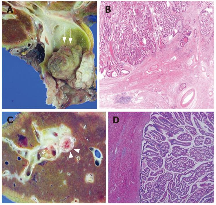
Figure 2 Macroscopic and microscopic findings of early bile duct cancers.
A, B: Extrahepatic early bile duct cancer. A papillary mass (arrows) protruding into the common bile duct lumen is noted (A). Although it is fairly large, its invasion is confined within the fibromuscular layer (HE, × 100) (B). C, D: An intrahepatic early bile duct cancer. Gross specimen of resected liver in patients with papillary mass (arrow heads) within the right intrahepatic bile duct. Tumor invasion is confined within the fibromuscular layer without hepatic parenchymal invasion (HE, × 10).
- Citation: Cha JM, Kim MH, Jang SJ. Early bile duct cancer. World J Gastroenterol 2007; 13(25): 3409-3416
- URL: https://www.wjgnet.com/1007-9327/full/v13/i25/3409.htm
- DOI: https://dx.doi.org/10.3748/wjg.v13.i25.3409


