Copyright
©2007 Baishideng Publishing Group Co.
World J Gastroenterol. Jun 28, 2007; 13(24): 3396-3399
Published online Jun 28, 2007. doi: 10.3748/wjg.v13.i24.3396
Published online Jun 28, 2007. doi: 10.3748/wjg.v13.i24.3396
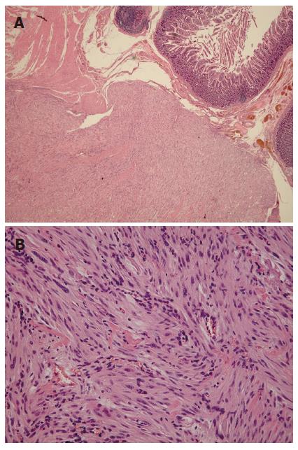
Figure 5 A well-defined subserosal duodenal GIST showing interlacing fascicles of the spindle cells with elongated cytoplasm (H&E stain; A: × 20, B: × 200).
- Citation: Kwon SH, Cha HJ, Jung SW, Kim BC, Park JS, Jeong ID, Lee JH, Nah YW, Bang SJ, Shin JW, Park NH, Kim DH. A gastrointestinal stromal tumor of the duodenum masquerading as a pancreatic head tumor. World J Gastroenterol 2007; 13(24): 3396-3399
- URL: https://www.wjgnet.com/1007-9327/full/v13/i24/3396.htm
- DOI: https://dx.doi.org/10.3748/wjg.v13.i24.3396


