Copyright
©2007 Baishideng Publishing Group Co.
World J Gastroenterol. Jun 28, 2007; 13(24): 3342-3349
Published online Jun 28, 2007. doi: 10.3748/wjg.v13.i24.3342
Published online Jun 28, 2007. doi: 10.3748/wjg.v13.i24.3342
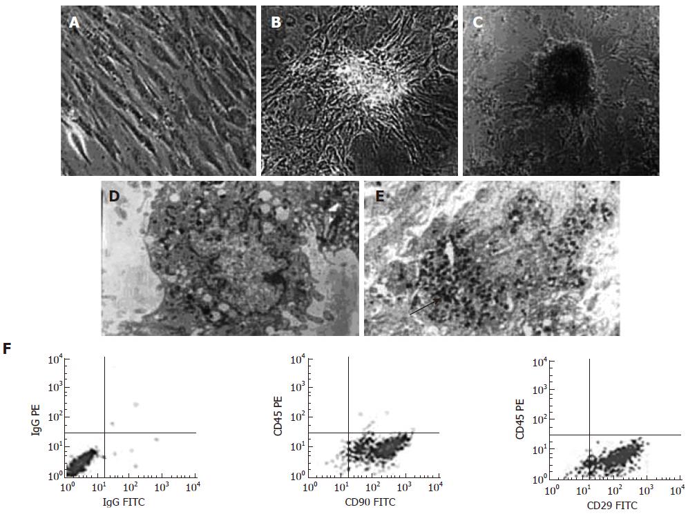
Figure 1 Morphological changes of BM-MSCs during differentiation.
A: The third passage of pre-induced BM-MSCs (× 500); B: BM-MSCs formed islet-like clusters under 5% FBS HG-DMEM culture (× 500); C: Some clusters were half suspended in the culture medium after induced by nicotinamide and exendin-4 (× 500); Electron microscopy: Secretory granules (the black arrow shows) are densely packed within the cytoplasm of D-MSCs (E, × 4000) whereas few is found in pre-induced BM-MSCs (D × 5000); F: Surface markers of BM-MSCs showed that CD90 and CD29 positive rates were more than 93% whereas CD45 expressions were less than 1%.
- Citation: Wu XH, Liu CP, Xu KF, Mao XD, Zhu J, Jiang JJ, Cui D, Zhang M, Xu Y, Liu C. Reversal of hyperglycemia in diabetic rats by portal vein transplantation of islet-like cells generated from bone marrow mesenchymal stem cells. World J Gastroenterol 2007; 13(24): 3342-3349
- URL: https://www.wjgnet.com/1007-9327/full/v13/i24/3342.htm
- DOI: https://dx.doi.org/10.3748/wjg.v13.i24.3342


