Copyright
©2007 Baishideng Publishing Group Co.
World J Gastroenterol. Jun 28, 2007; 13(24): 3311-3315
Published online Jun 28, 2007. doi: 10.3748/wjg.v13.i24.3311
Published online Jun 28, 2007. doi: 10.3748/wjg.v13.i24.3311
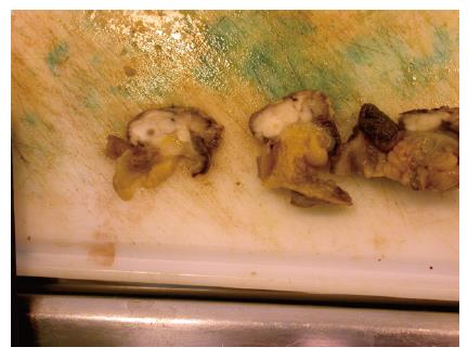
Figure 2 Cut surface of the leiomyosarcoma presented in Figure 1, showing a possible necrosis and a white, fish-flesh-like color, as typical for sarcomas.
The surface did not bulge on incision (Courtesy by S Duun).
- Citation: Ponsaing LG, Kiss K, Hansen MB. Classification of submucosal tumors in the gastrointestinal tract. World J Gastroenterol 2007; 13(24): 3311-3315
- URL: https://www.wjgnet.com/1007-9327/full/v13/i24/3311.htm
- DOI: https://dx.doi.org/10.3748/wjg.v13.i24.3311


