Copyright
©2007 Baishideng Publishing Group Co.
World J Gastroenterol. Jun 21, 2007; 13(23): 3262-3264
Published online Jun 21, 2007. doi: 10.3748/wjg.v13.i23.3262
Published online Jun 21, 2007. doi: 10.3748/wjg.v13.i23.3262
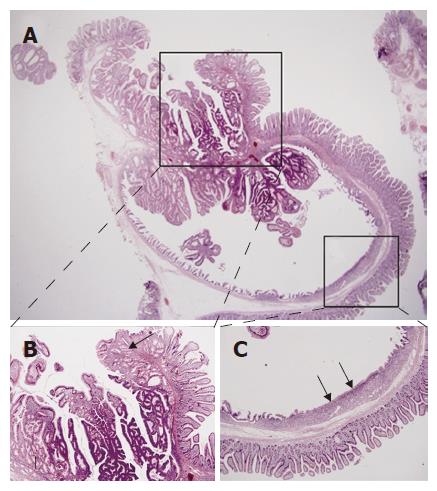
Figure 2 A: Inverted cystic lesion extends along the Brunner gland duct into the submucosa (HE, × 15); B: Normal or hyperplastic Brunner’s glands (arrows) are seen beneath the tubulovillous adenomatous lesion growing into the cyst lumen (HE, × 40); C: The flat surface epithelium and small mucus glands (arrows) are seen among the villiform epithelium (HE, × 40).
- Citation: Kim JH, Choi JW, Seo YS, Lee BJ, Yeon JE, Kim JS, Byun KS, Bak YT, Kim I, Park JJ. Inverted cystic tubulovillous adenoma involving Brunner’s glands of duodenum. World J Gastroenterol 2007; 13(23): 3262-3264
- URL: https://www.wjgnet.com/1007-9327/full/v13/i23/3262.htm
- DOI: https://dx.doi.org/10.3748/wjg.v13.i23.3262


