Copyright
©2007 Baishideng Publishing Group Co.
World J Gastroenterol. Jun 21, 2007; 13(23): 3176-3182
Published online Jun 21, 2007. doi: 10.3748/wjg.v13.i23.3176
Published online Jun 21, 2007. doi: 10.3748/wjg.v13.i23.3176
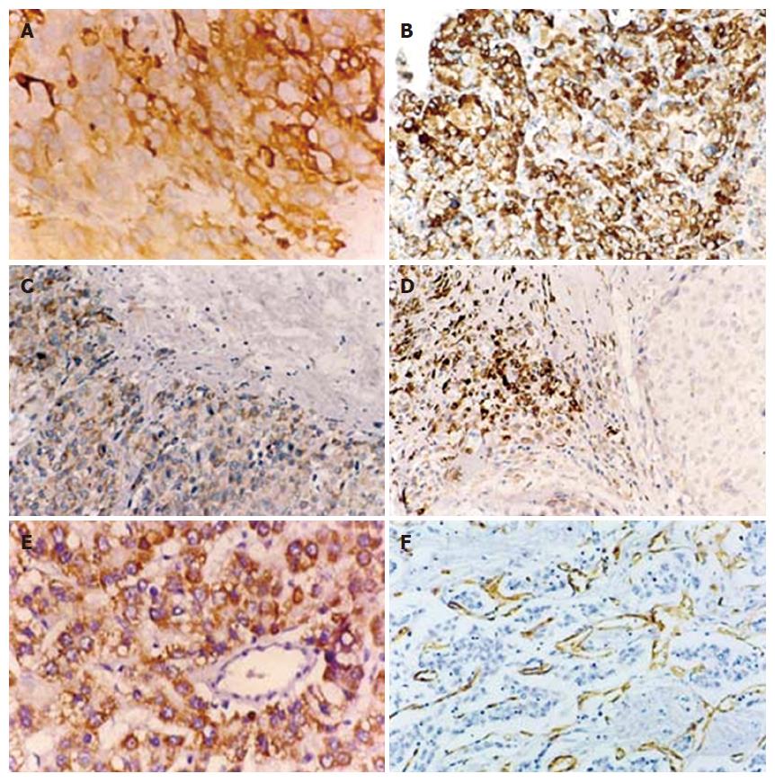
Figure 1 Representative examples of immunohistochemical staining for HIF-2α/EPASE1, VEGF, and CD31 in HCC.
A: Strong cytoplasmic immunore-activity of HIF-2α/EPASE1 is observed in cancers cells (× 400). B: Strong staining in the cytoplasm of noncancerous cirrhotic tissue (× 400). C: HIF-2α/EPAS1-positive staining in perinecrotic area (N) and D: In the cytoplasm of macrophages (× 400). E: Parallel studies of VEGF protein (cytoplasmic staining) and F: CD31 (for microvessels) immunohistochemistry performed on HCC (× 200).
- Citation: Bangoura G, Liu ZS, Qian Q, Jiang CQ, Yang GF, Jing S. Prognostic significance of HIF-2α/EPAS1 expression in hepatocellular carcinoma. World J Gastroenterol 2007; 13(23): 3176-3182
- URL: https://www.wjgnet.com/1007-9327/full/v13/i23/3176.htm
- DOI: https://dx.doi.org/10.3748/wjg.v13.i23.3176


