Copyright
©2007 Baishideng Publishing Group Co.
World J Gastroenterol. Jun 21, 2007; 13(23): 3153-3158
Published online Jun 21, 2007. doi: 10.3748/wjg.v13.i23.3153
Published online Jun 21, 2007. doi: 10.3748/wjg.v13.i23.3153
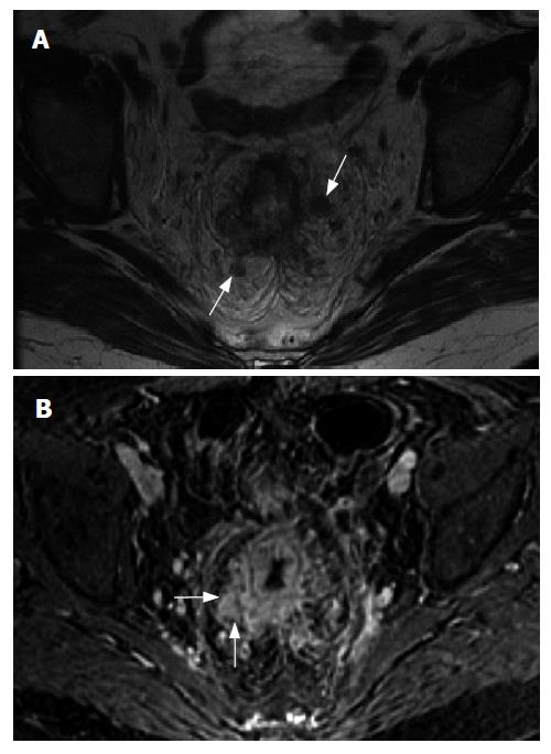
Figure 1 A 52 years old woman with rectal cancer.
Axial T2 (A) and axial fat suppressed gadolinium-enhanced T1-weighted (B) MR images demonstrate circumferential soft tissue thickening and abnormal enhancement of the rectum consistent with a neoplasm. Direct mesorectal invasion is present (arrows in B) as well as perirectal adenopathy (arrows in A).
- Citation: Berman L, Israel GM, McCarthy SM, Weinreb JC, Longo WE. Utility of magnetic resonance imaging in anorectal disease. World J Gastroenterol 2007; 13(23): 3153-3158
- URL: https://www.wjgnet.com/1007-9327/full/v13/i23/3153.htm
- DOI: https://dx.doi.org/10.3748/wjg.v13.i23.3153


