Copyright
©2007 Baishideng Publishing Group Co.
World J Gastroenterol. Jun 7, 2007; 13(21): 2932-2938
Published online Jun 7, 2007. doi: 10.3748/wjg.v13.i21.2932
Published online Jun 7, 2007. doi: 10.3748/wjg.v13.i21.2932
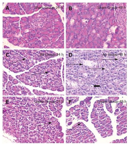
Figure 2 Light microscopy showing normal pancreatic tissue in the sham group (A, B) (HE, × 200), broad single cell necrosis (arrow) and significant increase in vacuolization (bold arrow) in the AP group at 24 h and 48 h (C, D) (HE, × 400), attenuated necrosis and vacuolization after leptin treatment at 24 h (E) (HE, × 400).
Mild edema was observed at 48 h in leptin-treated rats (F, arrow).
- Citation: Gultekin FA, Kerem M, Tatlicioglu E, Aricioglu A, Unsal C, Bukan N. Leptin treatment ameliorates acute lung injury in rats with cerulein-induced acute pancreatitis. World J Gastroenterol 2007; 13(21): 2932-2938
- URL: https://www.wjgnet.com/1007-9327/full/v13/i21/2932.htm
- DOI: https://dx.doi.org/10.3748/wjg.v13.i21.2932


