Copyright
©2007 Baishideng Publishing Group Co.
World J Gastroenterol. May 28, 2007; 13(20): 2775-2783
Published online May 28, 2007. doi: 10.3748/wjg.v13.i20.2775
Published online May 28, 2007. doi: 10.3748/wjg.v13.i20.2775
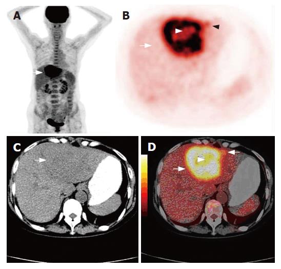
Figure 3 A 68-year-old female complaining of uncomfortable left upper abdomen for one month with increasing of CA19-9 (> 700 μ/L) (A), non-enhanced CT (C) detecting a low density lesion in the left lobe of the liver (white arrow), 18F-FDG PET (B) and fused imaging of PET/CT (D) showing necrosis (arrow head) in the center of the mass with a satellite lesion (black arrow).
CC was confirmed by liver biopsy.
- Citation: Sun L, Wu H, Guan YS. Positron emission tomography/computer tomography: Challenge to conventional imaging modalities in evaluating primary and metastatic liver malignancies. World J Gastroenterol 2007; 13(20): 2775-2783
- URL: https://www.wjgnet.com/1007-9327/full/v13/i20/2775.htm
- DOI: https://dx.doi.org/10.3748/wjg.v13.i20.2775


