Copyright
©2007 Baishideng Publishing Group Co.
World J Gastroenterol. May 28, 2007; 13(20): 2775-2783
Published online May 28, 2007. doi: 10.3748/wjg.v13.i20.2775
Published online May 28, 2007. doi: 10.3748/wjg.v13.i20.2775
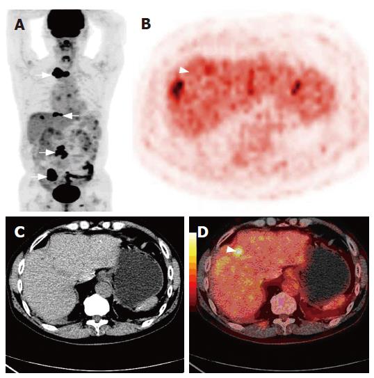
Figure 2 A 68-year-old male undergoing HCC resection 28-mo ago.
Multiples bone metastases (arrow) on the image of whole body PET (A), non-enhanced CT (C) detecting no recurrence lesion in the liver, 18F-FDG PET (B) and PET/CT fused imaging (D) showing a high metabolism recurrent lesion (arrow head).
- Citation: Sun L, Wu H, Guan YS. Positron emission tomography/computer tomography: Challenge to conventional imaging modalities in evaluating primary and metastatic liver malignancies. World J Gastroenterol 2007; 13(20): 2775-2783
- URL: https://www.wjgnet.com/1007-9327/full/v13/i20/2775.htm
- DOI: https://dx.doi.org/10.3748/wjg.v13.i20.2775


