Copyright
©2007 Baishideng Publishing Group Co.
World J Gastroenterol. May 28, 2007; 13(20): 2775-2783
Published online May 28, 2007. doi: 10.3748/wjg.v13.i20.2775
Published online May 28, 2007. doi: 10.3748/wjg.v13.i20.2775
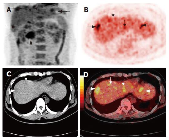
Figure 1 A 33-year-old man undergoing ascending colon cancer resection two years ago.
Coronal PET image (A) also showing recurrent lesion (arrow head) at the root of mesentery, 18F-FDG PET (B) and PET/CT fused imaging (D) demonstrating multiple hepatic metastases (arrow), and non-enhanced CT (C) detecting fewer lesions than PET/CT fused imaging (D).
- Citation: Sun L, Wu H, Guan YS. Positron emission tomography/computer tomography: Challenge to conventional imaging modalities in evaluating primary and metastatic liver malignancies. World J Gastroenterol 2007; 13(20): 2775-2783
- URL: https://www.wjgnet.com/1007-9327/full/v13/i20/2775.htm
- DOI: https://dx.doi.org/10.3748/wjg.v13.i20.2775


