Copyright
©2007 Baishideng Publishing Group Co.
World J Gastroenterol. Jan 14, 2007; 13(2): 306-309
Published online Jan 14, 2007. doi: 10.3748/wjg.v13.i2.306
Published online Jan 14, 2007. doi: 10.3748/wjg.v13.i2.306
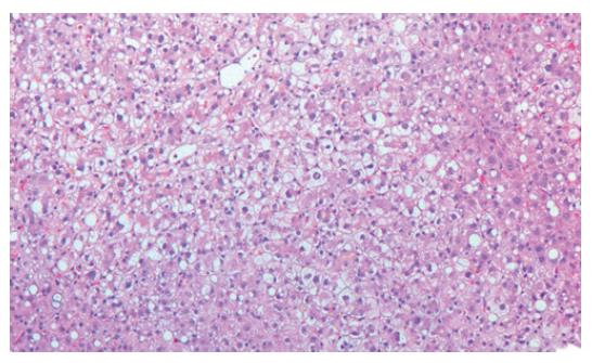
Figure 2 HE-staining of a liver biopsy derived from a 23-yr-old male presenting with liver failure after exertional heat stroke.
Microscopy showed liver tissue with intact architecture of the lobules. Portal fields showed no signs of fibrosis or inflammatory infiltration. Accentuated centrolobular necrosis was accompanied with fatty degeneration of hepatocytes. Additionally, focal bile inclusions were found without Mallory bodies, iron debris and atypical cellular proliferation. Altogether, the morphological picture does not fit acute Wilson’s disease, hemochromatosis or infection, but ischemic liver disease.
- Citation: Weigand K, Riediger C, Stremmel W, Flechtenmacher C, Encke J. Are heat stroke and physical exhaustion underestimated causes of acute hepatic failure? World J Gastroenterol 2007; 13(2): 306-309
- URL: https://www.wjgnet.com/1007-9327/full/v13/i2/306.htm
- DOI: https://dx.doi.org/10.3748/wjg.v13.i2.306


