Copyright
©2007 Baishideng Publishing Group Co.
World J Gastroenterol. Jan 14, 2007; 13(2): 192-218
Published online Jan 14, 2007. doi: 10.3748/wjg.v13.i2.192
Published online Jan 14, 2007. doi: 10.3748/wjg.v13.i2.192
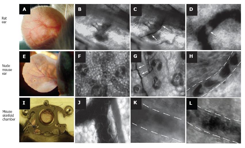
Figure 1 Label-free imaging of blood vessels in different animal models.
Rat ear (with hair): A: External view of large vessel; B, C: transmission images of microvessel at low magnification (4 ×) before (B) and after (C) topical administration of an optical clearing agent such as glycerol (arrows show microvessel); D: high-resolution image of rolling WBC (arrow) in a venula (magnification 40 ×). Ear of nude mouse: E: external view of large vessels; F, G: transmission images of microvessels at low magnification (10 ×) before (F) and after (G) topical administration of glycerol (arrows show microvessel); H: high-resolution image of individual RBCs in a capillary (magnification 100 ×); I: large vessels in skinfold chamber of a mouse; J: transmission image of blood microvessel at low magnification (10 ×); K, I: high-resolution image of a venula before (K) and after (I) topical administration of glycerol (40 ×).
-
Citation: Galanzha EI, Tuchin VV, Zharov VP. Advances in small animal mesentery models for
in vivo flow cytometry, dynamic microscopy, and drug screening. World J Gastroenterol 2007; 13(2): 192-218 - URL: https://www.wjgnet.com/1007-9327/full/v13/i2/192.htm
- DOI: https://dx.doi.org/10.3748/wjg.v13.i2.192


