Copyright
©2007 Baishideng Publishing Group Co.
World J Gastroenterol. May 21, 2007; 13(19): 2758-2760
Published online May 21, 2007. doi: 10.3748/wjg.v13.i19.2758
Published online May 21, 2007. doi: 10.3748/wjg.v13.i19.2758
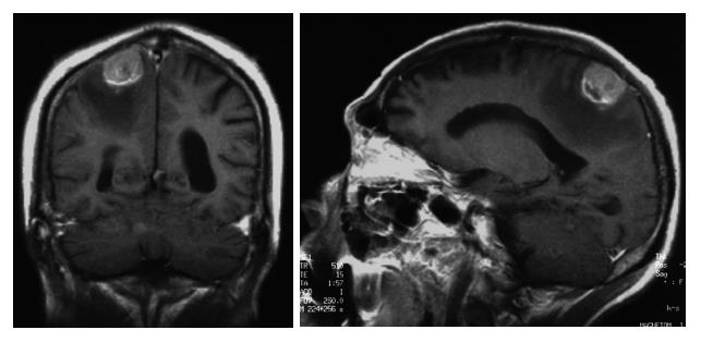
Figure 3 Head MRI.
A 2.5-cm tumor heterogeneously enhanced on contrast imaging in the parietal region. A low-intensity area, is an edematous change, extended from the right parietal region to the frontal lobe white matter.
- Citation: Tamaki K, Shimizu I, Urata M, Kohno N, Fukuno H, Ito S, Sano N. A patient with spinal metastasis from hepatocellular carcinoma discovered from neurological findings. World J Gastroenterol 2007; 13(19): 2758-2760
- URL: https://www.wjgnet.com/1007-9327/full/v13/i19/2758.htm
- DOI: https://dx.doi.org/10.3748/wjg.v13.i19.2758


