Copyright
©2007 Baishideng Publishing Group Co.
World J Gastroenterol. May 21, 2007; 13(19): 2675-2683
Published online May 21, 2007. doi: 10.3748/wjg.v13.i19.2675
Published online May 21, 2007. doi: 10.3748/wjg.v13.i19.2675
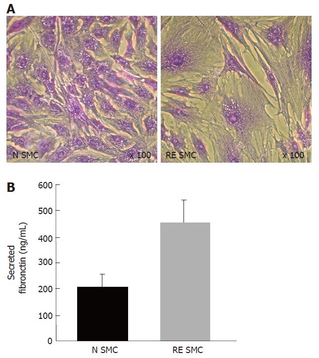
Figure 3 A: Bright field photomicrograph of fibrosis-derived intestinal smooth muscle cells (RE SMC) and normal cells (N SMC) observed after crystal violet staining; B: Fibronectin secretion level in RE SMC and N SMC assessed by ELISA (Chemicon).
- Citation: Haydont V, Vozenin-Brotons MC. Maintenance of radiation-induced intestinal fibrosis: Cellular and molecular features. World J Gastroenterol 2007; 13(19): 2675-2683
- URL: https://www.wjgnet.com/1007-9327/full/v13/i19/2675.htm
- DOI: https://dx.doi.org/10.3748/wjg.v13.i19.2675


