Copyright
©2007 Baishideng Publishing Group Co.
World J Gastroenterol. May 21, 2007; 13(19): 2649-2654
Published online May 21, 2007. doi: 10.3748/wjg.v13.i19.2649
Published online May 21, 2007. doi: 10.3748/wjg.v13.i19.2649
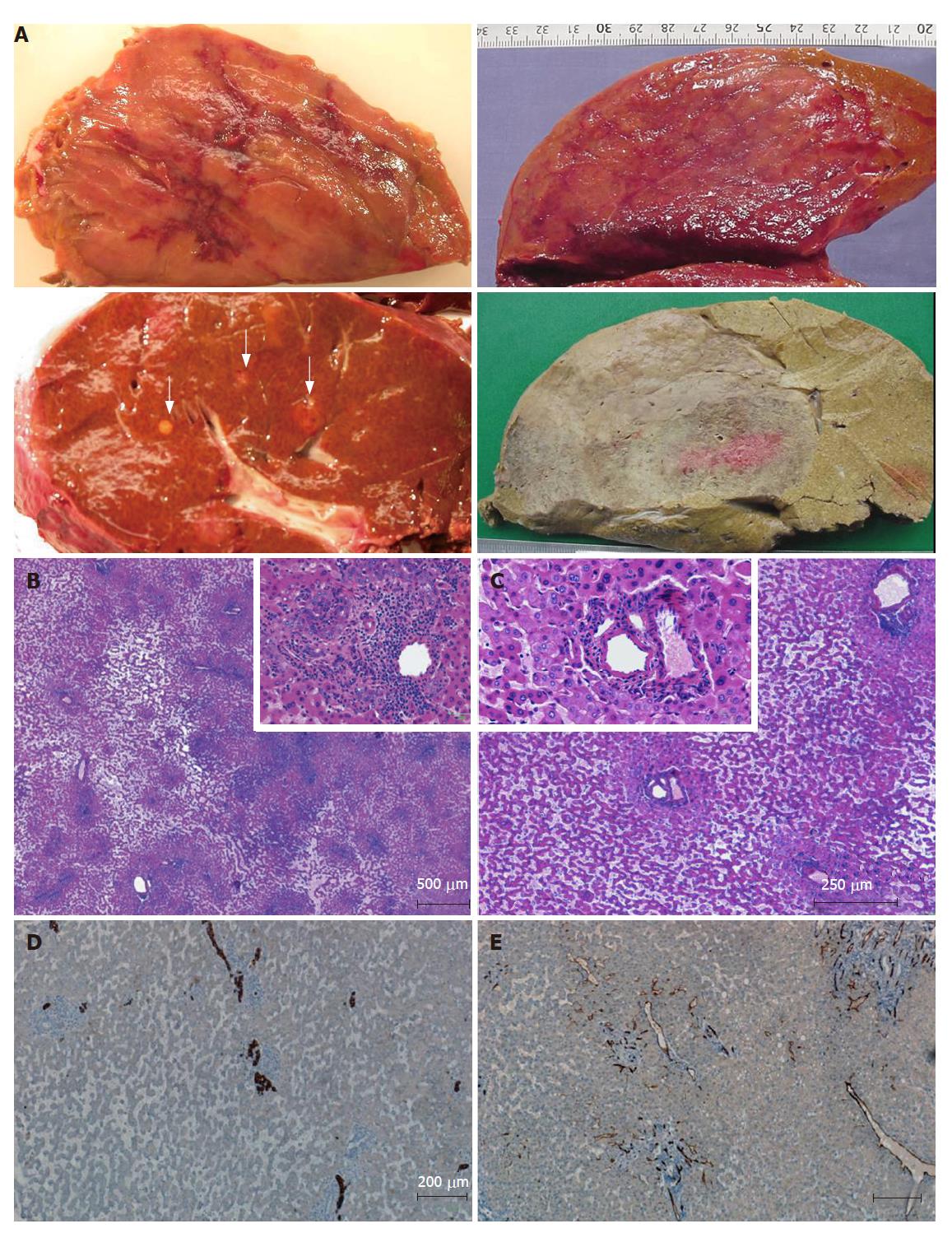
Figure 3 Inflammatory/telangiectatic adenomas (from different cases).
A: several sections of adenomas showing different aspects. Upper left: hemorrhagic areas in the center of the tumor; lower left: several micronodules (white arrows) in a patient with a large inflammatory/telangiectatic adenoma (not shown); upper right: this massive adenoma is difficult to identify on fresh tissue; the same is more easily identified after fixation (lower right); B, C and insets: typical aspects with obvious telangiectasia and inflammatory infiltrates (B) around thick arteries (C) (HE); D: some ductular reaction is visible on cytokeratin 7 immunostaining; E: CD34 immunostaining highlights capillarized sinusoids around arteries.
- Citation: Bioulac-Sage P, Blanc JF, Rebouissou S, Balabaud C, Zucman-Rossi J. Genotype phenotype classification of hepatocellular adenoma. World J Gastroenterol 2007; 13(19): 2649-2654
- URL: https://www.wjgnet.com/1007-9327/full/v13/i19/2649.htm
- DOI: https://dx.doi.org/10.3748/wjg.v13.i19.2649


