Copyright
©2007 Baishideng Publishing Group Co.
World J Gastroenterol. May 21, 2007; 13(19): 2649-2654
Published online May 21, 2007. doi: 10.3748/wjg.v13.i19.2649
Published online May 21, 2007. doi: 10.3748/wjg.v13.i19.2649
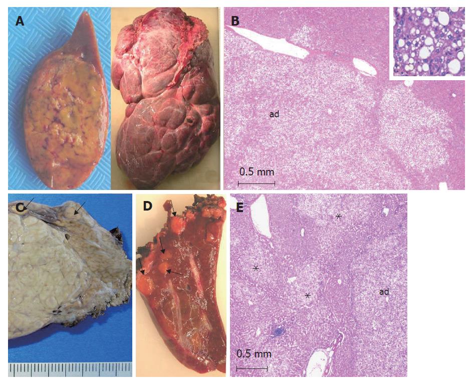
Figure 1 HNF1α-mutated adenomas (from different cases).
A: left: single, yellowish HCA; right: a massive form involving the whole right lobe made of several numerous adjacent nodules of different sizes; B: typical aspect of a steatotic HCA (ad) with a lobulated contour. Inset: HCA at a higher magnification showing macro and microvesicular steatosis (HE); C: left: part of a large focal nodular hyperplasia (indication of surgery), nearby small adenomas (arrows) discovered by chance on the surface of the liver (corresponding to a somatic HNF1α-mutated adenomatosis). D: several yellow microadenomas in a somatic HNF1α-mutated adenomatosis; E: multiple not well limited steatotic microadenomas (asterix) nearby a larger adenoma (ad) in a patient with a constitutional HNF1α-mutated adenomatosis.
- Citation: Bioulac-Sage P, Blanc JF, Rebouissou S, Balabaud C, Zucman-Rossi J. Genotype phenotype classification of hepatocellular adenoma. World J Gastroenterol 2007; 13(19): 2649-2654
- URL: https://www.wjgnet.com/1007-9327/full/v13/i19/2649.htm
- DOI: https://dx.doi.org/10.3748/wjg.v13.i19.2649


