Copyright
©2007 Baishideng Publishing Group Co.
World J Gastroenterol. Apr 28, 2007; 13(16): 2385-2387
Published online Apr 28, 2007. doi: 10.3748/wjg.v13.i16.2385
Published online Apr 28, 2007. doi: 10.3748/wjg.v13.i16.2385
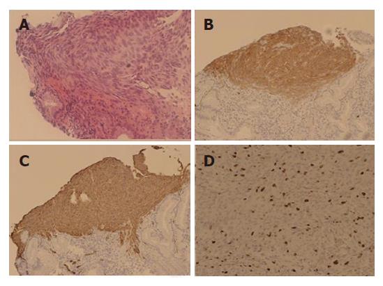
Figure 4 Photomicrographs of biopsy specimens from the fistula showing spindle-shaped cells (A) (HE, × 200), immunohistochemical staining showing a diffuse positive signal for KIT (B) (× 100), a diffuse positive signal for CD34 (C) (× 100), and MIB-1 (D).
The labeling index (L.I) was 18% (× 200).
- Citation: Osada T, Nagahara A, Kodani T, Namihisa A, Kawabe M, Yoshizawa T, Ohkusa T, Watanabe S. Gastrointestinal stromal tumor of the stomach with a giant abscess penetrating the gastric lumen. World J Gastroenterol 2007; 13(16): 2385-2387
- URL: https://www.wjgnet.com/1007-9327/full/v13/i16/2385.htm
- DOI: https://dx.doi.org/10.3748/wjg.v13.i16.2385


