Copyright
©2007 Baishideng Publishing Group Co.
World J Gastroenterol. Apr 28, 2007; 13(16): 2385-2387
Published online Apr 28, 2007. doi: 10.3748/wjg.v13.i16.2385
Published online Apr 28, 2007. doi: 10.3748/wjg.v13.i16.2385
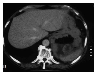
Figure 3 Enhanced com-puted tomography (CT) scan of the abdomen revealing a large central cavity in the tumor partially filled with fluid, and an irregular thickening of the gastric wall in the posterior aspect of the fundus extending into the left subphrenic space.
- Citation: Osada T, Nagahara A, Kodani T, Namihisa A, Kawabe M, Yoshizawa T, Ohkusa T, Watanabe S. Gastrointestinal stromal tumor of the stomach with a giant abscess penetrating the gastric lumen. World J Gastroenterol 2007; 13(16): 2385-2387
- URL: https://www.wjgnet.com/1007-9327/full/v13/i16/2385.htm
- DOI: https://dx.doi.org/10.3748/wjg.v13.i16.2385


