Copyright
©2007 Baishideng Publishing Group Co.
World J Gastroenterol. Apr 21, 2007; 13(15): 2243-2246
Published online Apr 21, 2007. doi: 10.3748/wjg.v13.i15.2243
Published online Apr 21, 2007. doi: 10.3748/wjg.v13.i15.2243
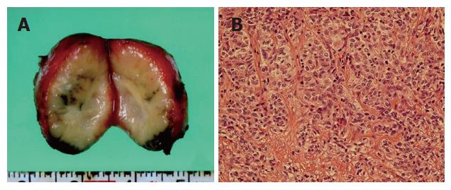
Figure 5 A well-capsulated solid mass at the cut surface (A) and a well-capsulated solid lymph node containing moderately differentiated adenocarcinoma similar in appearance to the pathology of the primary tumor (B) in the specimen of the patient's liver.
- Citation: Ito Y, Tajima Y, Fujita F, Tsutsumi R, Kuroki T, Kanematsu T. Solitary recurrence of hilar cholangiocarcinoma in a mediastinal lymph node two years after curative resection. World J Gastroenterol 2007; 13(15): 2243-2246
- URL: https://www.wjgnet.com/1007-9327/full/v13/i15/2243.htm
- DOI: https://dx.doi.org/10.3748/wjg.v13.i15.2243


