Copyright
©2007 Baishideng Publishing Group Co.
World J Gastroenterol. Apr 21, 2007; 13(15): 2160-2165
Published online Apr 21, 2007. doi: 10.3748/wjg.v13.i15.2160
Published online Apr 21, 2007. doi: 10.3748/wjg.v13.i15.2160
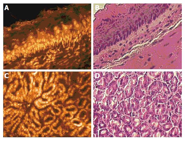
Figure 1 A: Esophagus after intravenous injection of acriflavine hydrochloride: The germinative zone and the columnar arrangement within the layered structure can be easily visualized.
Note that the mouse esophagus is keratinized; B: Ex vivo H&E staining of the corresponding specimen; C: In vivo visualization of gastric glands. Note the dos-à-dos-arrangement of the glands. Only scarce nuclei stain within the loose connective tissue of the lamina propria; D: Corresponding specimen of the mouse stomach in a transverse section.
-
Citation: Goetz M, Memadathil B, Biesterfeld S, Schneider C, Gregor S, Galle PR, Neurath MF, Kiesslich R.
In vivo subsurface morphological and functional cellular and subcellular imaging of the gastrointestinal tract with confocal mini-microscopy. World J Gastroenterol 2007; 13(15): 2160-2165 - URL: https://www.wjgnet.com/1007-9327/full/v13/i15/2160.htm
- DOI: https://dx.doi.org/10.3748/wjg.v13.i15.2160


