Copyright
©2007 Baishideng Publishing Group Co.
World J Gastroenterol. Apr 14, 2007; 13(14): 2077-2082
Published online Apr 14, 2007. doi: 10.3748/wjg.v13.i14.2077
Published online Apr 14, 2007. doi: 10.3748/wjg.v13.i14.2077
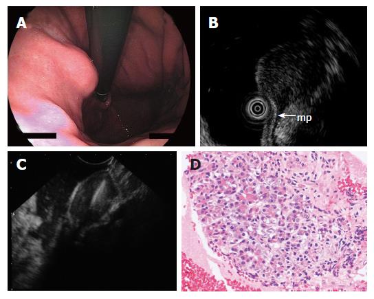
Figure 5 A: Endoscopy showing submucosal lesion in the upper body of the stomach; B: EUS using ultrasound catheter probe demonstrating 1.
5 cm subepithelial hypoechoic tumor with continuity to proper muscle layer (arrow-mp); C: The hypoechoic mass shown on EUS was punctured under real-time EUS guidance; D: The FNAB specimen consisted of acinar cells. The histologic diagnosis was ectopic pancreas.
- Citation: Akahoshi K, Sumida Y, Matsui N, Oya M, Akinaga R, Kubokawa M, Motomura Y, Honda K, Watanabe M, Nagaie T. Preoperative diagnosis of gastrointestinal stromal tumor by endoscopic ultrasound-guided fine needle aspiration. World J Gastroenterol 2007; 13(14): 2077-2082
- URL: https://www.wjgnet.com/1007-9327/full/v13/i14/2077.htm
- DOI: https://dx.doi.org/10.3748/wjg.v13.i14.2077


