Copyright
©2007 Baishideng Publishing Group Co.
World J Gastroenterol. Apr 14, 2007; 13(14): 2061-2065
Published online Apr 14, 2007. doi: 10.3748/wjg.v13.i14.2061
Published online Apr 14, 2007. doi: 10.3748/wjg.v13.i14.2061
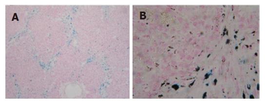
Figure 5 Iron deposition in rat liver induced by DMN (Prussian blue).
A: model group A, the iron is mainly accompanied by fiber (× 100); B: model group B, similar to the model A in iron deposition, iron nodules are deep blue in color and mainly located extracellularly or in the Kupffer cells (× 400).
- Citation: He JY, Ge WH, Chen Y. Iron deposition and fat accumulation in dimethylnitrosamine-induced liver fibrosis in rat. World J Gastroenterol 2007; 13(14): 2061-2065
- URL: https://www.wjgnet.com/1007-9327/full/v13/i14/2061.htm
- DOI: https://dx.doi.org/10.3748/wjg.v13.i14.2061


