Copyright
©2007 Baishideng Publishing Group Co.
World J Gastroenterol. Apr 14, 2007; 13(14): 2061-2065
Published online Apr 14, 2007. doi: 10.3748/wjg.v13.i14.2061
Published online Apr 14, 2007. doi: 10.3748/wjg.v13.i14.2061
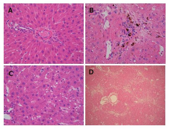
Figure 2 Liver histopathology of DMN-treated rats (HE).
A: Control group, no marked pathological changes (× 400); B: Group model A, hemorrhagic necrosis with foci of lymphomonocytic infiltration around fibrosis tissue can be seen (× 400); C: Group model B, fat accumulated in numerous liver cells (× 400); D: Group model B, quantities of fiber deposited and linked with each other (× 400).
- Citation: He JY, Ge WH, Chen Y. Iron deposition and fat accumulation in dimethylnitrosamine-induced liver fibrosis in rat. World J Gastroenterol 2007; 13(14): 2061-2065
- URL: https://www.wjgnet.com/1007-9327/full/v13/i14/2061.htm
- DOI: https://dx.doi.org/10.3748/wjg.v13.i14.2061


