Copyright
©2007 Baishideng Publishing Group Co.
World J Gastroenterol. Apr 7, 2007; 13(13): 1995-1997
Published online Apr 7, 2007. doi: 10.3748/wjg.v13.i13.1995
Published online Apr 7, 2007. doi: 10.3748/wjg.v13.i13.1995
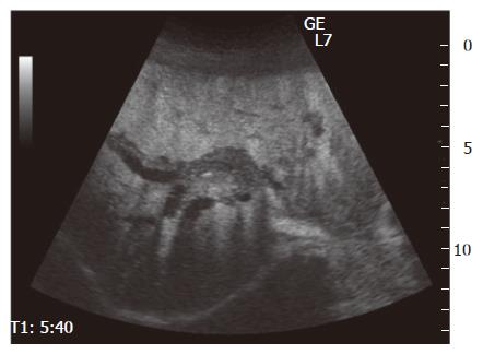
Figure 1 Contrast-enhanced ultrasonography (CEUS) showing thickened wall of intrahepatic bile ducts (from hilar to peripheral) with dilation, and the lesion was well enhanced.
- Citation: Matsumoto N, Yokoyama K, Nakai K, Yamamoto T, Otani T, Ogawa M, Tanaka N, Iwasaki A, Arakawa Y, Sugitani M. A case of eosinophilic cholangitis: Imaging findings of contrast-enhanced ultrasonography, cholangioscopy, and intraductal ultrasonography. World J Gastroenterol 2007; 13(13): 1995-1997
- URL: https://www.wjgnet.com/1007-9327/full/v13/i13/1995.htm
- DOI: https://dx.doi.org/10.3748/wjg.v13.i13.1995


