Copyright
©2007 Baishideng Publishing Group Co.
World J Gastroenterol. Apr 7, 2007; 13(13): 1912-1927
Published online Apr 7, 2007. doi: 10.3748/wjg.v13.i13.1912
Published online Apr 7, 2007. doi: 10.3748/wjg.v13.i13.1912
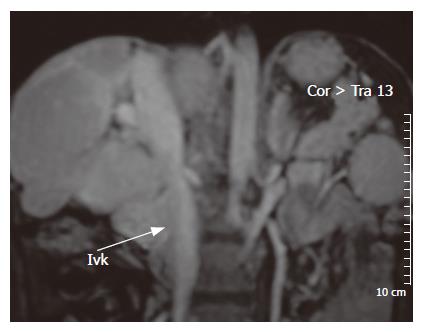
Figure 6 Magnetic resonance imaging of the liver with contrasting agent shows that inferior vena cava is compressed by hypertrophy of caudate lobe and gross lobulation of the liver.
- Citation: Bayraktar UD, Seren S, Bayraktar Y. Hepatic venous outflow obstruction: Three similar syndromes. World J Gastroenterol 2007; 13(13): 1912-1927
- URL: https://www.wjgnet.com/1007-9327/full/v13/i13/1912.htm
- DOI: https://dx.doi.org/10.3748/wjg.v13.i13.1912


