Copyright
©2007 Baishideng Publishing Group Co.
World J Gastroenterol. Mar 21, 2007; 13(11): 1752-1754
Published online Mar 21, 2007. doi: 10.3748/wjg.v13.i11.1752
Published online Mar 21, 2007. doi: 10.3748/wjg.v13.i11.1752
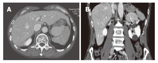
Figure 3 An abdominal contrast-enhanced CT.
A: A well-marginated ovoid mass, approximately 6 cm in diameter; B: Supplied by a vascular branch arising from the splenic artery. There are renal cysts.
- Citation: Chin S, Isomoto H, Mizuta Y, Wen CY, Shikuwa S, Kohno S. Enlarged accessory spleen presenting stomach submucosal tumor. World J Gastroenterol 2007; 13(11): 1752-1754
- URL: https://www.wjgnet.com/1007-9327/full/v13/i11/1752.htm
- DOI: https://dx.doi.org/10.3748/wjg.v13.i11.1752


