Copyright
©2007 Baishideng Publishing Group Co.
World J Gastroenterol. Mar 21, 2007; 13(11): 1652-1658
Published online Mar 21, 2007. doi: 10.3748/wjg.v13.i11.1652
Published online Mar 21, 2007. doi: 10.3748/wjg.v13.i11.1652
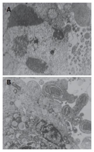
Figure 3 Morphology of HepG2 treated with UDCA (0.
4 mmol/L) for 48 h (TEM, × 4000). A: Nuclear frag-mentation,chromosome condensation, cell shrinkage and loss of cell-cell contact are visible; B: Subsequent ruffling and blebbling of the cell membrane, and formation of apoptotic bodies are also observed.
- Citation: Liu H, Qin CY, Han GQ, Xu HW, Meng M, Yang Z. Mechanism of apoptotic effects induced selectively by ursodeoxycholic acid on human hepatoma cell lines. World J Gastroenterol 2007; 13(11): 1652-1658
- URL: https://www.wjgnet.com/1007-9327/full/v13/i11/1652.htm
- DOI: https://dx.doi.org/10.3748/wjg.v13.i11.1652


