Copyright
©2007 Baishideng Publishing Group Co.
World J Gastroenterol. Mar 14, 2007; 13(10): 1632-1635
Published online Mar 14, 2007. doi: 10.3748/wjg.v13.i10.1632
Published online Mar 14, 2007. doi: 10.3748/wjg.v13.i10.1632
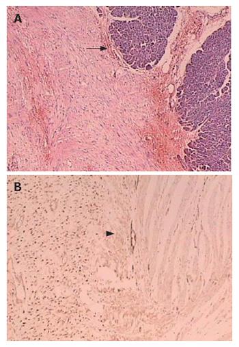
Figure 5 Tumoral lesion consisted of spindle cells growing in sweeping fascicles, with eosinophilic cytoplasm and occasional focal infiltration of plump nuclei in pancreas (arrow) (A) and bowel wall (arrow head) (B).
Most of the tumor cells showed immunoreactivity for β-catenin (+++).
- Citation: Sun L, Wu H, Zhuang YZ, Guan YS. A rare case of pregnancy complicated by mesenteric mass: What does chylous ascites tell us? World J Gastroenterol 2007; 13(10): 1632-1635
- URL: https://www.wjgnet.com/1007-9327/full/v13/i10/1632.htm
- DOI: https://dx.doi.org/10.3748/wjg.v13.i10.1632


