Copyright
©2007 Baishideng Publishing Group Co.
World J Gastroenterol. Mar 14, 2007; 13(10): 1505-1515
Published online Mar 14, 2007. doi: 10.3748/wjg.v13.i10.1505
Published online Mar 14, 2007. doi: 10.3748/wjg.v13.i10.1505
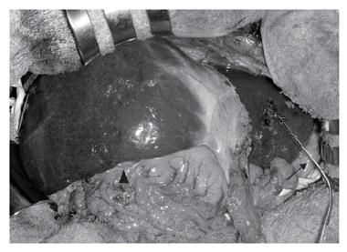
Figure 5 Intraoperative view at laparotomy after biliary drainage and portal vein embolization.
A percutaneous transhepatic biliary drainage tube (arrow) has been inserted into the bile duct of segment 3. The right liver is markedly atrophic, and there is a clear line of demarcation between the right and left liver.
- Citation: Seyama Y, Makuuchi M. Current surgical treatment for bile duct cancer. World J Gastroenterol 2007; 13(10): 1505-1515
- URL: https://www.wjgnet.com/1007-9327/full/v13/i10/1505.htm
- DOI: https://dx.doi.org/10.3748/wjg.v13.i10.1505


